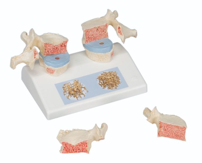
Anatomical Model: Osteoporosis Model This magnetic osteoporosis model offers a side-by-side comparison of healthy vs. osteoporotic thoracic vertebrae using anatomically accurate reproductions of T11 and T12. Designed for visual and tactile learning, the model features two osteoporotic vertebrae with narrowed intervertebral discs on the left and two healthy vertebrae with full discs on the right.
The included base features a detailed 3D illustration with micro CT imagery from real bone biopsies, providing additional insight into structural changes caused by osteoporosis at the microscopic level.
Key Features:
Magnetic display of T11 and T12 vertebrae
Side-by-side comparison of osteoporotic vs. healthy bone
Shows narrowing of intervertebral discs and bone density loss
Ideal for patient education and clinical demonstrations
Includes base with 3D micro CT images for added context
Compact and portable—perfect for desktops, clinics, or classrooms
Digital Learning Bonus: Every 3B Scientific Anatomy model includes access to the 3B Smart Anatomy app. Register your product to:
Extend your warranty from 3 to 5 years
Get 1 year of free access to premium digital content:
23 anatomy lectures
117 interactive 3D models
39 quizzes for enhanced learning
Perfect for healthcare professionals, educators, and osteoporosis awareness programs. Clearly demonstrate the effects of bone degeneration—with hands-on visuals and digital tools.


 Palmer's Cocoa Butter Formula Lotion with Vitamin E - 7.25 oz Jar